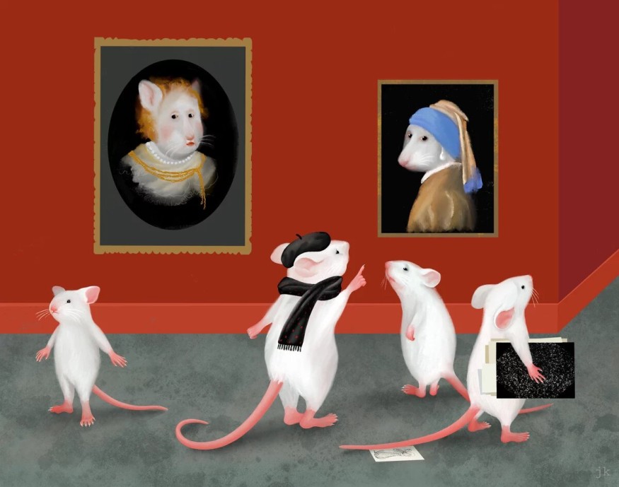
Some painting creations made by Rembrandt and Vermeer are identical. Most of the time, people make mistakes in crediting one artwork to the other painter. However, identifying the different compositions and details makes experts see the difference between the masterpieces made by the two great artists.
Max Planck Institute of Neurobiology recently uncovered how the ability needed on this specific challenge could be applied. With the help of mice subjects, researchers knew how the brain differentiates images into categories. The research determined that these categories are found in the lobes where visualization is processed. The findings helped the expert understand how knowledge and semantic memories are stored across a functional brain.
How Does Semantic Memory Work?

Art professionals hone their expertise by including a habit of differentiating multiple artworks made available by various painters. Through this process, they can develop a sense of distinction in one artwork over another. Like many other repetitive functions, this process is acquired through knowledge that is being gathered with the use of semantic memory.
Semantic memory is a type of long-term recall procured not by specific experience but by gaining exposure before a piece of abstract information. However, the details regarding this type of memory and its storage in the human brain have limited studies and supporting evidence. Max Planck's neurobiologist, expert, and author of the study Pieter Golstein said in a Florida News Time report that understanding how semantic memories are stored requires them to specify first the right area of the brain where it is stored. The study was made possible with the help of mice subjects taught how to acquire the skill and complex knowledge that art experts have.
ALSO READ : Brain Cells Destroyed as Neurons Learn Faster; DNA Breaks May Provide Answer to Genetics
Where is Semantic Memory Stored?
The study involved experiments that included tasks similar to the complex ability of art professionals. In the experiment, the mice subjects were exposed to different images that contained stripe patterns. However, these patterns have different appearances on each of the images. The experiment's goal was for the subjects to learn how these stripe images vary from each other. Throughout the examination, the mice did not experience any difficulties sorting the stripe images based on their categories. The quick-learning ability of the subjects has allowed them to store category features on their brain. However, when the stripe images were moved to a different location, the mouse was confused. This led the scientists to identify that the category learning is indeed heavily linked with the visual cortex.
The examination also included the tracking of neuron activities as the subjects learned throughout the experiment. According to the study, the learning process allowed more regions of the visual cortex to develop category skills. Neurobiology expert and lead author of the study Mark Hübener said that the neurons located at the visual cortex could receive two distinct signals: the visual stimulus and the category information based on what they saw on the stripe images. The study was published in the journal Nature Neuroscience, titled "Mouse visual cortex areas represent perceptual and semantic features of learned visual categories."
RELATED ARTICLE : Humanized Lab-Grown 'Mini-Brains' Imitates Parkinson's Disease and Dementia; May Replace Mouse Models in Neurological Studies
Check out more news and information on Neurology in Science Times.


