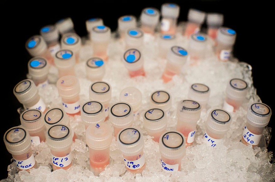
Thanks to 'virtual biopsies' incorporating CT and ultrasound scans, cancer patients may one day be able to forgo intrusive tissue sampling.
Experts from Cambridge have demonstrated that routine diagnostic scans may build a visual tumor guide to help doctors find the right place for targeted biopsies.
Although also enabling extensive sampling of a tumor, this may allow less intrusive biopsies and even make physical biopsies redundant one day.
University of Cambridge radiologist Lucian Beer said their research is a move forward in unraveling tumor heterogeneity non-invasively. For ultrasound-guided targeted biopsies, they used standard-of-care CT-based radiomic tumor environments.
Specialists reported the full conclusions of the research in the journal European Radiology.
What is Tumor Heterogeneity?
Tumor heterogeneity is the concept provided by doctors that various cell types are made up of a given lump of cancerous tissues.
Understanding the structure of a given tumor is crucial to choosing the patient's right medication since genetically diverse cells may vary in their treatment responses.
Therefore, cancer patients usually receive various biopsies to validate their diagnose and help inform their care strategy - with physicians juggling the desire to take detailed samples alongside the painful aspect of the operation.
Accurate biopsies are particularly relevant in the case of ovarian cancer, the researchers clarified. The most popular form appears to come with elevated tumor heterogeneity levels,' high grade serous ovarian cancer.'
Unfortunately, those patients with greater heterogeneity levels within their forms of tumor cells appear to have weaker treatment regime responses.
Dr. Beer and colleagues enrolled six advanced ovarian cancer patients in their research who were expected to have ultrasound-guided biopsies before beginning their chemotherapy regimen.
The team used regular CT scans to create a three-dimensional representation of patients' tumors, made up from a sequence of X-ray photographs' slice-by-slice.'
The researchers then used high-powered computational methods to chart the tumor's magnitude and internal characteristics, which were subsequently superimposed on the ultrasound scan to direct each case's biopsies.
How Simulated Biopsies Could Help Therapeutic Biopsies In The Future

According to the team, the method effectively captured the variety of cancer cells inside each patient's tumor.
The researchers said that simulated biopsies might supplement intrusive therapeutic biopsies in the future.
Professor Evis Sala of the Department of Radiology has suggested that this research is a significant landmark in tissue sampling quality. The experts say that in turning cutting edge science into standard clinical treatment, they are testing the limits.
This feasibility analysis includes researchers from the radiology school, the Cambridge Institute of Cancer Treatment UK, the NHS Foundation Trust of Cambridge University Hospitals, and more.
ALSO READ : How Binge Drinking Affects Your Brain Activity
Check out more news and information on Medicine and Health on Science Times.


