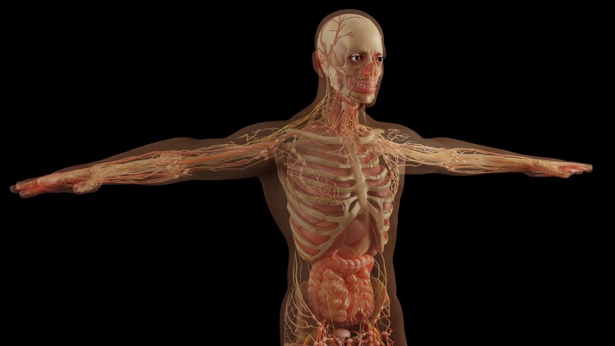
Scripps Research Institute scientists recently announced a new tissue-clearing technique that renders biological samples transparent. The said novel technology makes it easier and faster for scientists to study healthy and diseased organs of the body.
The new method combines the elements of two prior approaches in tissue-clearing technology and is more practical and scalable than either of them. Neuroscience assistant professor Li Ye, Ph. D. from Scripps Research said that this new method is a simple and universal tissue-clearing technique for large-sample applications, even on animals.

Developing the Tissue-Clearing Technique
It is critical to image intact tissues to gain a complete understanding of the biological processes in health and disease, according to a paper published on the website of the University of Rochester Medical Center. But it is not easy due to the non-uniform light absorption, scattering, attenuation patterns, and refractive index (RI) of organs.
Tissue-clearing techniques will make biological samples transparent by homogenizing the RIs of the tissues. It is achieved by replacing or dissolving molecules with different RIs through chemical processes. It is typically combined with imaging techniques to study the cellular structures and organizations of intact tissue.
According to Interesting Engineering's report, the novel tissue-clearing technique started 15 years ago and is still under development. The idea started because researchers wanted to trace nerve connections within whole brains.
However, the technology only worked for brains and not other organs or body parts, likely because the methods included either organic or water-based solvents that may cause complications.
Organic solvents reduce the production of fluorescent signals that could tell the active genes or molecules that are of interest in the study. Meanwhile, the water-based solvents were impractically weak to be used in tissue clearing of other organs. More so, these techniques use labor-intensive and often hazardous chemicals.
ALSO READ : Atlas of Cranial Stem Cells May Give Insights to Normal Head Development, Craniofacial Birth Defects
HYBRiD: Hydrogel-Based Reinforcement DISCO
Due to the challenges scientists faced from previous experiments, they decided to improve the method. Ye, the co-first author of the paper, said that previous methods could not be used in an ordinary lab and at a large scale. But the new method devised by scientists at Scripps Research Institue bypasses those issues.
Dubbed HYBRiD (hydrogel-based reinforcement of three-dimensional imaging solvent-cleared organs (DISCO)), this new method employs water-based hydrogels to protect molecules within the tissues needed to be preserved.
Researchers recombined components of organic and polymer-based clearing pipelines and were able to achieve high transparency and protein retention, according to the news release in EurekAlert! More so, they were able to bring about compatibility with direct fluorescent imaging and immunostaining in SARS-CoV-2-infected cells in the whole chest of their mice models for the first time.
Study co-first author Victoria Nudell, a research assistant at Ye's lab, said that this novel tissue-clearing technique makes it practical and scalable for routine use in an ordinary lab.
They published the full findings of their study, titled "HYBRiD: Hydrogel-Reinforced DISCO for Clearing Mammalian Bodies," in the journal Nature Methods.
RELATED ARTICLE: Genome Editing for Microbiomes Available Soon Through New CRISPR Development
Check out more news and information on Biology in Science Times.


